
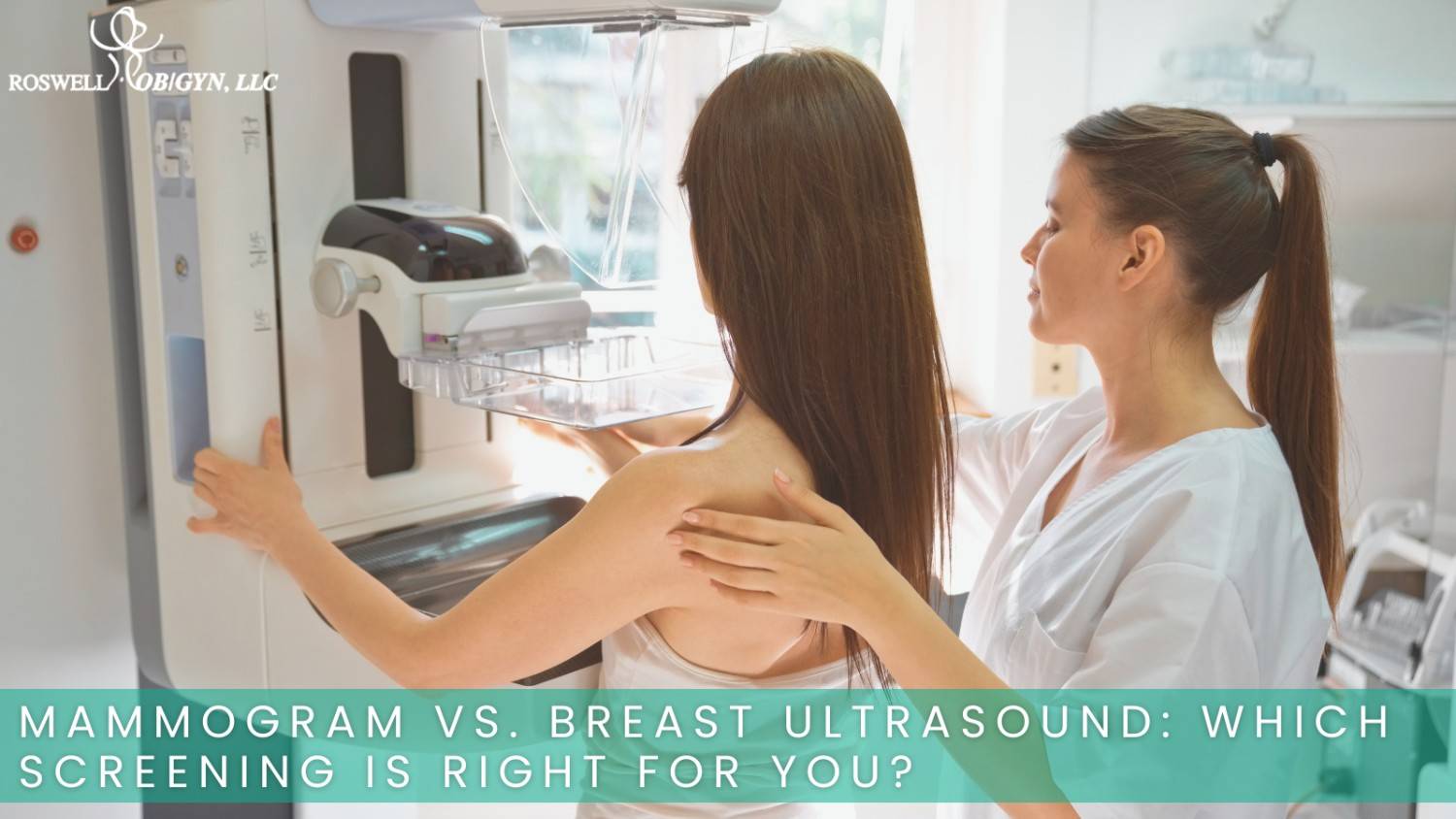
A mammogram and a breast ultrasound are not competing tests, they are complementary tools that work together to protect your breast health. A mammogram uses low-dose X-rays and remains the gold-standard screening for detecting early breast cancer, especially in women age 40 and older. A breast ultrasound uses sound waves and is most often used as a follow-up diagnostic test or as a supplemental screening for women with dense breast tissue. Our practice provides both imaging options as part of our breast imaging and mammography services, and your provider will recommend the right test based on your age, risk profile, and breast density. Understanding what each screening does, and what it cannot do, helps you make confident decisions about your care.
How a Mammogram Works
A mammogram is a low-dose X-ray that captures detailed images of breast tissue. During the exam, each breast is briefly compressed between two plates so the radiologist can see fine structures clearly. The compression usually lasts only a few seconds per image and feels firm rather than painful for most patients.
Mammograms are the primary tool for routine breast cancer screening because they can find disease years before a lump becomes large enough to feel. The exam is especially effective at picking up:
- Microcalcifications, the tiny calcium deposits that can be one of the earliest signs of cancer
- Very small tumors, including stage 0 (ductal carcinoma in situ) cancers
- Changes in tissue structure that appear normal on physical exam
- Asymmetries between the two breasts that may need closer review
Standard 2D mammography and 3D mammography (tomosynthesis) both use the same X-ray principle, with 3D capturing multiple thin slices that give radiologists a clearer view of overlapping tissue. You can read more about advanced imaging on our 3D mammography page and learn how it fits alongside other common screening tests.
How a Breast Ultrasound Works
A breast ultrasound uses high-frequency sound waves, not radiation, to produce real-time images of breast tissue. A small handheld probe is moved across the breast, and the echoes are converted into pictures on a monitor. The exam takes 15 to 30 minutes, is painless, and requires no compression or special preparation.
Ultrasound is most useful when your provider needs to take a closer look at a specific area. It is particularly good at:
- Distinguishing a fluid-filled cyst from a solid mass after a lump is found on exam or mammogram
- Imaging dense breast tissue, which can hide tumors on a mammogram
- Guiding a needle during a biopsy procedure
- Evaluating breast changes in women under 30 or in patients who are pregnant or breastfeeding
The trade-off is that ultrasound cannot see microcalcifications as clearly as a mammogram, which is why it almost always works alongside mammography rather than replacing it. For pregnancy-related ultrasound services, see our obstetric ultrasound page.
ALSO READ: 3D Mammogram vs. 2D Mammogram: Is the Upgrade Worth It?
Mammogram vs. Breast Ultrasound: Quick Comparison
Both tests image breast tissue, but they answer different clinical questions and rely on completely different technology. The table below summarizes the key differences at a glance, including how each exam works, who it best serves, and the role it plays in your overall breast health plan. Use it as a reference when discussing your screening options with your provider.
| Feature | Mammogram | Breast Ultrasound |
| Technology | Low-dose X-ray | High-frequency sound waves |
| Radiation | Yes, very low dose | None |
| Best for | Routine screening, microcalcifications | Cysts, dense tissue, biopsy guidance |
| Typical age group | 40 and older for routine screening | Any age, often used under 30 |
| Compression | Yes | No |
| Detects microcalcifications | Yes | Limited |
| Detects cancer in dense breasts | Less effective | More effective as a supplement |
| Role | Primary screening | Diagnostic and supplemental |
When a Mammogram Is the Right Choice
A mammogram is the recommended screening for most women starting at age 40, and earlier for those with a personal or family history of breast cancer. Major organizations including the American Cancer Society and the American College of Obstetricians and Gynecologists support annual or biennial mammograms beginning at 40, with personalized screening for higher-risk patients starting as early as 30.
You are likely a candidate for a routine mammogram if:
- You are 40 or older with average risk
- You have a family history of breast or ovarian cancer
- You carry a known BRCA1, BRCA2, or other high-risk genetic variant
- You have had previous breast biopsies showing certain high-risk findings
- Your provider has recommended annual imaging based on your overall risk profile
Even if you have dense breasts, the mammogram remains the foundation of your screening plan. A supplemental ultrasound or MRI may be added on top, not used in place of it. Patients planning a family-history review may also benefit from a visit through our broader gynecological services.
When a Breast Ultrasound Makes More Sense
A breast ultrasound is most appropriate when there is a specific question that needs answering, or when your breast tissue makes a mammogram harder to read. Your provider may order one in the following situations:
- You feel a new lump, area of thickening, or focal pain
- A recent mammogram showed an area that needs further evaluation
- You have dense breast tissue noted on your last screening
- You are under age 30 with a palpable breast concern
- You are pregnant or breastfeeding and need imaging
- A biopsy is planned and the radiologist needs real-time guidance
Ultrasound is also helpful for women who cannot have a mammogram for medical reasons, though this is rare. In almost every other case, a breast ultrasound supports the mammogram rather than replacing it.
ALSO READ: What to Expect During a Mammogram: First-Timer’s Guide
What Dense Breast Tissue Means for Your Screening
Roughly 40 to 50 percent of women have dense breast tissue, which means a higher proportion of glandular and fibrous tissue compared with fat. Dense tissue appears white on a mammogram, and so do tumors, which can make small cancers harder to see.
If your mammography report says you have heterogeneously dense or extremely dense breasts, your provider may suggest adding a breast ultrasound or breast MRI to your annual screening. This combined approach can find cancers that a mammogram alone might miss, while still using the mammogram as the primary tool for microcalcifications and early-stage disease. Federal guidelines now require mammography facilities in the United States to notify patients about their breast density, so you will receive this information in writing after your screening.
How to Choose the Right Screening for You
Your ideal screening plan depends on three main factors: your age, your breast density, and your personal risk for breast cancer. A simple way to think about it:
- Routine screening at age 40 or older: Start with a mammogram
- Dense breast tissue: Combine a mammogram with a supplemental breast ultrasound or MRI
- New lump, pain, or visible change: Begin with a diagnostic mammogram, ultrasound, or both depending on age
- Under 30 with a breast concern: Start with an ultrasound, since young breast tissue is denser
- High genetic or family risk: Discuss earlier and more frequent screening with your provider, sometimes including breast MRI
Your provider will weigh these factors with you and recommend the imaging sequence that gives you the most useful information.
Schedule Your Breast Screening at Roswell Ob/Gyn, LLC
Your screening plan should match your age, breast density, and personal risk, not a one-size-fits-all schedule. The team at Roswell Ob/Gyn, LLC provides in-office mammography services and breast ultrasound across our Alpharetta, Atlanta, Canton, and Cumming locations, with personalized recommendations rooted in your full medical history. Whether you are due for a routine screening, following up on a recent finding, or have a new concern you want evaluated, our providers can guide you to the right imaging plan.
Request your appointment online or call our office today to schedule your mammogram, breast ultrasound, or combined screening with a provider who knows you, not just your file.
Frequently Asked Questions
Is a mammogram or breast ultrasound more accurate?
For routine cancer screening in average-risk women aged 40 and older, a mammogram is more accurate because it detects microcalcifications and very early tumors that ultrasound cannot see clearly. A breast ultrasound is more accurate for evaluating a specific lump, distinguishing cysts from solid masses, and screening dense breast tissue when used as a supplement.
Do I need both a mammogram and a breast ultrasound?
Most women do not need both every year. A routine mammogram is enough for average-risk patients. Your provider may add a breast ultrasound if you have dense breast tissue, a palpable lump, or an area on your mammogram that requires closer evaluation.
Does a breast ultrasound use radiation?
No. A breast ultrasound uses sound waves, not X-rays, and involves no radiation. That is why it is safe for women who are pregnant, breastfeeding, or under age 30, where minimizing radiation exposure is preferred.
At what age should I start getting a mammogram?
Most women with average risk should begin routine mammogram screening at age 40. Women with a family history of breast cancer, a known genetic mutation, or other risk factors may start earlier, sometimes at age 30 or even younger, based on individual evaluation by their provider.
Can a breast ultrasound replace my mammogram if I have dense breasts?
No. Even with dense breasts, a breast ultrasound is added to your mammogram, not used in place of it. The mammogram finds microcalcifications and structural changes that ultrasound can miss, while ultrasound helps see through dense tissue. Together they offer the most complete picture.